Art-labeling activity gross anatomy of the spinal cord embarks on a captivating journey, inviting readers to explore the intricate complexities of the spinal cord’s structure through an engaging and interactive approach.
This comprehensive guide delves into the external and internal anatomy of the spinal cord, unraveling the mysteries of its gray and white matter. It illuminates the intricate organization of the spinal cord into segments, providing a foundational understanding of this vital structure.
Gross Anatomy of the Spinal Cord
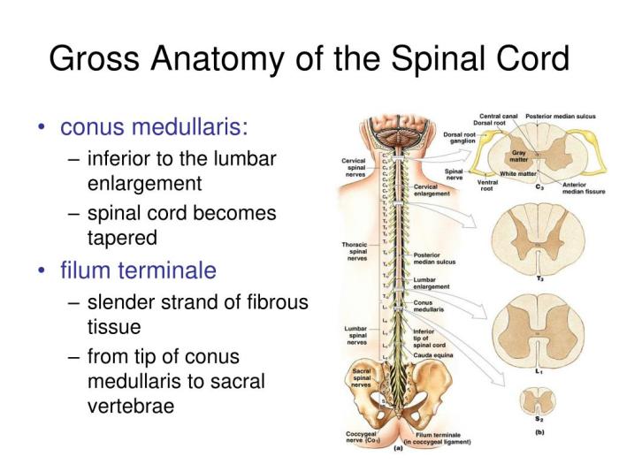
The spinal cord is a cylindrical structure that extends from the medulla oblongata in the brainstem to the lumbar region of the vertebral column. It is approximately 45 cm long in adults and has a diameter of about 1 cm.
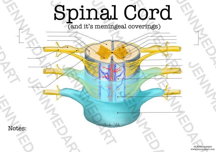
The spinal cord is surrounded by three layers of meninges: the dura mater, arachnoid mater, and pia mater.The external anatomy of the spinal cord can be divided into two regions: the cervical and lumbar enlargements. The cervical enlargement is located at the level of the C4-T1 vertebrae and contains the nerve roots that innervate the upper limbs.
The lumbar enlargement is located at the level of the L1-S3 vertebrae and contains the nerve roots that innervate the lower limbs.The internal anatomy of the spinal cord can be divided into two regions: the gray matter and the white matter.
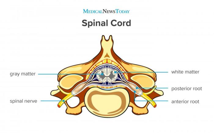
The gray matter is located in the center of the spinal cord and contains the cell bodies of neurons. The white matter is located on the outside of the gray matter and contains the axons of neurons.The spinal cord is organized into segments.
Each segment contains a pair of dorsal and ventral nerve roots. The dorsal nerve roots contain sensory neurons that transmit sensory information from the body to the spinal cord. The ventral nerve roots contain motor neurons that transmit motor information from the spinal cord to the muscles.
Art-Labeling Activity

Art-labeling activities are a great way to teach students about the gross anatomy of the spinal cord. To create an art-labeling activity, first create a diagram of the spinal cord. Then, create a table that lists the anatomical structures of the spinal cord and their corresponding labels.
Finally, provide instructions for students to label the structures on the diagram.Here is an example of an art-labeling activity that you can use to teach students about the gross anatomy of the spinal cord:Materials:* Diagram of the spinal cord
- Table of anatomical structures and their corresponding labels
- Colored pencils or markers
Instructions:
- Look at the diagram of the spinal cord and identify the different anatomical structures.
- Use the table to find the labels for the anatomical structures.
- Use colored pencils or markers to label the anatomical structures on the diagram.
Assessment:You can assess students’ understanding of the gross anatomy of the spinal cord by having them complete the art-labeling activity. You can also ask students to write a short essay describing the different anatomical structures of the spinal cord.
Examples and Methods
Art-labeling activities can be used in a variety of ways to teach the gross anatomy of the spinal cord. Here are a few examples:*
-*As a pre-lab activity
Students can complete an art-labeling activity before they dissect a spinal cord. This will help them to familiarize themselves with the different anatomical structures of the spinal cord.
-
-*As a review activity
Students can complete an art-labeling activity after they have dissected a spinal cord. This will help them to reinforce their understanding of the different anatomical structures of the spinal cord.
-*As an assessment tool
Students can complete an art-labeling activity as an assessment of their understanding of the gross anatomy of the spinal cord.
Here are a few tips for creating effective art-labeling activities:*
- *Use clear and concise instructions. Students should be able to easily understand what they are supposed to do.
- *Provide a variety of resources. Students should have access to a variety of resources, such as diagrams, tables, and textbooks, to help them complete the activity.
- *Make the activity engaging. Students are more likely to learn if they are engaged in the activity. Try to make the activity fun and interesting.
Assessment and Evaluation: Art-labeling Activity Gross Anatomy Of The Spinal Cord

Art-labeling activities can be used to assess students’ understanding of the gross anatomy of the spinal cord. Here is a rubric that you can use to evaluate students’ art-labeling activities:Criteria:*
-*Accuracy
The student correctly labels all of the anatomical structures on the diagram.
-
-*Completeness
The student labels all of the anatomical structures on the diagram.
-*Neatness
The student’s labels are neat and legible.
Scoring:*
-*4 points
The student meets all of the criteria.
-
-*3 points
The student meets most of the criteria.
-*2 points
The student meets some of the criteria.
-*1 point
The student meets few of the criteria.
-*0 points
The student does not meet any of the criteria.
Art-labeling activities are a valuable assessment tool because they allow students to demonstrate their understanding of the gross anatomy of the spinal cord in a creative way.
Illustrations and Images

-*Illustration 1
This illustration shows the external anatomy of the spinal cord. The spinal cord is surrounded by three layers of meninges: the dura mater, arachnoid mater, and pia mater. The spinal cord is divided into two regions: the cervical and lumbar enlargements.Illustration
2:This illustration shows the internal anatomy of the spinal cord. The gray matter is located in the center of the spinal cord and contains the cell bodies of neurons. The white matter is located on the outside of the gray matter and contains the axons of neurons.Illustration
3:This illustration shows the organization of the spinal cord into segments. Each segment contains a pair of dorsal and ventral nerve roots. The dorsal nerve roots contain sensory neurons that transmit sensory information from the body to the spinal cord.
The ventral nerve roots contain motor neurons that transmit motor information from the spinal cord to the muscles.
Q&A
What are the benefits of using art-labeling activities in teaching gross anatomy?
Art-labeling activities enhance visual memory, promote active learning, and facilitate deeper comprehension of anatomical structures.
How can I create an effective art-labeling activity?
Design clear and accurate diagrams, provide concise labels, and incorporate opportunities for students to test their understanding.
How can I assess students’ understanding through art-labeling activities?
Develop a rubric that evaluates the accuracy and completeness of students’ labeling, as well as their understanding of the anatomical concepts.